Images
Images are from WikiMedia Commons and the Public Health Image Library (PHIL).

|
Afr Tryp Life Cycle African trypanosome life cycle Permissions: Public domain http://commons.wikimedia.org/wiki/File:AfrTryp_LifeCycle.gif |

|
Afric tryp 1a DPDxi Two areas from a blood smear from a patient with African trypanosomiasis (sleeping sickness). Thin blood smear stained with Giemsa. Typical trypomastigote stages (the only stages found in patients), with a posterior kinetoplast, a centrally located nucleus, an undulating membrane, and an anterior flagellum. The two Trypanosoma brucei species that cause human trypanosomiasis, T. b. gambiense and T. b. rhodesiense, are indistinguishable morphologically. Permissions: Public domain http://commons.wikimedia.org/wiki/File:Afric_tryp_1a_DPDxi.jpg |

|
Babesia equi Permissions: GNU Free Documentation License http://commons.wikimedia.org/wiki/File:Babesia_equi.jpg |

|
Babesia life cycle Babesia life cycle Permissions: Public domain http://commons.wikimedia.org/wiki/File:Babesia_LifeCycle.gif |
|
|
Babesia life cycle human Life cycle of the Parasite Babesia, (B.microti or B.divergens) including the infection to humans. Permissions: GNU Free Documentation License http://commons.wikimedia.org/wiki/File:Babesia_life_cycle_human_en.svg |

|
Babesien Hund Babesia infected red blood cell in a dog Permissions: Public domain http://commons.wikimedia.org/wiki/File:Babesien_Hund.jpg |
|
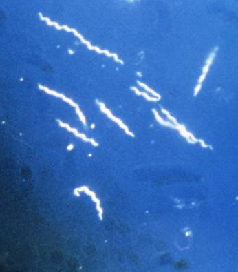
Borrelia burgdorferi Using darkfield microscopy technique, this photomicrograph, magnified 400x, reveals the presence of spirochete, or ?corkscrew-shaped? bacteria known as Borrelia burgdorferi, which is the pathogen responsible for causing Lyme disease. Borrelia burgdorferi are helical shaped bacteria, and are about 10-25Ám long. These bacteria are transmitted to humans by the bite of an infected deer tick, and caused more than 23,000 cases of Lyme disease in the United States during 2002. Permissions: Public domain http://commons.wikimedia.org/wiki/File:Borrelia_burgdorferi-cropped.jpg |
|
|
Borrelia drawing Borrelia drawing (Borrelia burgdorferi) ; Borrelia is a genus of bacteria of the spirochete phylum. It causes borreliosis, a zoonotic, vector-borne disease transmitted primarily by ticks and some by lice, depending on the species. There are 36 known species of Borrelia. Permissions: GNU Free Documentation License http://commons.wikimedia.org/wiki/File:BorreliaDrawing.jpg |

|
Borrelia life cycle Permissions: Public domain http://commons.wikimedia.org/wiki/File:Borrelia_cycle.jpg |
|
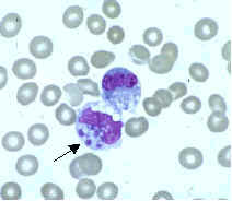
Echaff Ehrlichia chaffeensis primarily infects mononuclear leukocytes (predominantly monocytes and macrophages), but may also be seen occasionally in the granulocytes of some patients with severe disease. Permissions: Public domain http://commons.wikimedia.org/wiki/File:Echaff.jpg |

|
Ehrlichia drawing Infection of a cell by Ehrlicha. 1: bacteria entering in cell. 2, 3, 4 : reproduction and Morula in cytoplasm of a neutrophil Permissions: GNU Free Documentation License, http://commons.wikimedia.org/wiki/File:EhrlichiaDrawing.jpg |

|
Erythema migrans - erythematous rash in Lyme disease - PHIL 9875 This 2007 photograph depicts the pathognomonic erythematous rash in the pattern of a ?bull?s-eye?, which manifested at the site of a tick bite on this Maryland woman?s posterior right upper arm, who?d subsequently contracted Lyme disease. Permissions: Public domain http://commons.wikimedia.org/wiki/File:Erythema_migrans_-_erythematous_rash_in_Lyme_disease_-_PHIL_9875.jpg |

|
Influenza virus particle color This (Pseudocolored) negative-stained (false-colored) transmission electron micrograph (TEM) depicts the ultrastructural details of an influenza virus particle, or ?virion?. A member of the taxonomic family Orthomyxoviridae, the influenza virus is a single-stranded RNA organism. Permissions: PD-USGov-HHS-CDC (None - This image is in the public domain and thus free of any copyright restrictions http://commons.wikimedia.org/wiki/File:Influenza_virus_particle_color.jpg |

|
Neisseria gonorrhoeae PHIL 3693 lores This illustration depicts a urethral exudate containing Neisseria gonorrhoeae from a patient with gonococcal urethritis. This illustration depicts a Gram-stain of a urethral exudate showing typical intracellular gram-negative diplococci, and pleomorphic extracellular gram-negative organisms, which is diagnostic for gonococcal urethritis Permissions: PD-USGov-HHS-CDC (None - This image is in the public domain and thus free of any copyright restrictions http://commons.wikimedia.org/wiki/File:Neisseria_gonorrhoeae_PHIL_3693_lores.jpg |
|
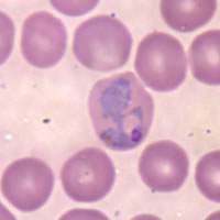
P vivax trophozoite4 Large, amoeboid trophozoites of P. vivax in a thin blood smear. Permissions: Public domain http://commons.wikimedia.org/wiki/File:P_vivax_trophozoite4.jpg |

|
PCP X-Ray Lung X-ray of patient shows infection with Pneumocystis carinii pneumonia. Permissions: Public domain http://commons.wikimedia.org/wiki/File:PCPxray.jpg |

|
Plasmodium lifecycle PHIL 3405 lores This is an illustration of the life cycle of the parasites of the genus Plasmodium that are causal agents of Malaria. Permissions: Public domain http://commons.wikimedia.org/wiki/File:Plasmodium_lifecycle_PHIL_3405_lores.jpg |

|
Plasmodium vivax Plasmodium vivax Permissions: Public domain http://commons.wikimedia.org/wiki/File:Plasmodium_vivax.jpg |
|
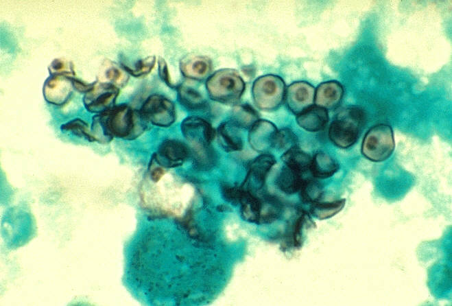
Pneumocystis carinii in smear from BAL 01ee046 lores Cysts of Pneumocystis carinii in smear from bronchoalveolar lavage. Methenamine silver stain. Permissions: Public domain http://commons.wikimedia.org/wiki/File:Pneumocystis_carinii_in_smear_from_BAL_01ee046_lores.jpg |
|
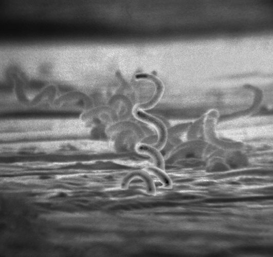
Treponema pallidum Electron micrograph of Treponema pallidum on cultures of cotton-tail rabbit epithelium cells (Sf1Ep). Treponema pallidum is the causative agent of syphilis. In the United States, over 35,600 cases of syphilis were reported by health officials in 1999. Permissions: Public domain http://commons.wikimedia.org/wiki/File:Treponema_pallidum.jpg |

|
Trypanosoma sp. PHIL 613 lores Trypanosoma forms in blood smear from patient with African trypanosomiasis. Trypanosoma forms in blood smear from patient with African trypanosomiasis. Parasite. Permissions: Public domain http://commons.wikimedia.org/wiki/File:Trypanosoma_sp._PHIL_613_lores.jpg |
Bioinformatics Center,
Institute for Chemical Research,
Kyoto University, Uji, Kyoto 611-0011, Japan
System last updated Thu, January 24, 2013 14:25:29 JST / SVN revision: 1054
Username: 17721B12964F44E75B28BFFCDA4FEE0B
